TidalHealth


Medical Services


Get access to your medical receipts and make payments for your medical bills.
Login to your patient portal to see all of your health records and messages.
Learn about open positions at TidalHealth and start down the path to your new career today.
“Our children are the best gift that we have ever received and the friendly compassionate and professional staff at TidalHealth’s Mother and Baby unit made it all the more easy and enjoyable.” Rob Gordy, Salisbury, MD
“My breast cancer diagnosis couldn’t have come at a worse time as the world was shutting down due to COVID-19. Friends were surprised to hear I wasn’t going across the bay for treatment. My doctors were all knowledgeable, professional, and compassionate.” Patti Weeg, Salisbury, MD
According to Dr. John Mansueti, executive medical director of cancer services for TidalHealth's Richard A. Henson Cancer Institute and TidalHealth's Allen Cancer Center, it takes a village to provide the...
In this episode of TidalHealth On Point, Tiffany Travers, LCSW-C, director of Behavioral Health Outpatient Services, and Stacey Walker, MSW, LCSW-C, clinical manager of Crisis Services, discuss TidalHealth's behavioral health...
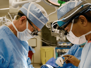
For 10 years in a row, TidalHealth has been named by Healthgrades as one of America's 50 Best Hospitals for Vascular Surgery. In this episode of TidalHealth on Point, vascular...
"It is health that is real wealth and not pieces of gold and silver." Mahatma Gandhi